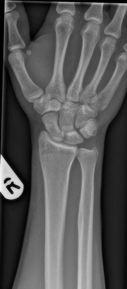
ap wrist | wrist ap position ap wrist This article provides a comprehensive approach to wrist radiographs, including techniques and common indications for imaging.
Assessing left ventricular systolic function. Section Progress. 0% Complete. Methods for assessing systolic function (contractile function) Several echocardiographic measurements are available to assess left ventricular systolic function.
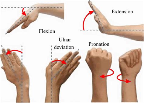
0 · wrist radial and ulnar deviation
1 · wrist ap view
2 · wrist ap position
3 · wrist ap lateral
4 · pa vs ap wrist xray
5 · normal ap wrist
6 · lateral view of the wrist
7 · ap wrist watch price
Since it can't do just one block, it increases it by the LP size, typically 4, 8, or 16 MB (depends on the size of your disk). To do that you can run the command : chfs -a size=+1 /usr. this will increase the size of the filesystem by one LP.
Although performed PA the view can often be referred to an AP view. Indications. The PA wrist radiograph is requested for myriad reasons including but not limited to trauma, . Wrist radiographic positioning tips for radiologic techs. Read about correct positions and projections used to obtain wrist radiographs.
borsa da lavoro gucci
aim at mid-carpus + 10° cephalad. Indications. alignment of bones/joints. carpal/lunate instability. Critique. volar cortex of pisiform + capitate within central 1/3 of volar . Learn how to position and expose the wrist for PA and AP x-ray views, and how to interpret the radiographic criteria. See examples of fractures, osteomyelitis, and arthritis in the wrist.The antero posterior or AP view or supinated view has its specific applications in evaluating wrist pathology.
This article provides a comprehensive approach to wrist radiographs, including techniques and common indications for imaging.
bob chapeau gucci
Posterior Anterior View of the Wrist. - See: - AP view. - Carpal height & Carpal height ratio. - Clenched Fist AP. - Radial Inclination. It is the orthogonal projection of the PA wrist. Indications. The lateral wrist radiograph is requested for myriad reasons including but not limited to trauma, suspected . What are the 2 key x-ray signs we should look for on the AP and the 2 key x-ray signs we should look for on the lateral for every wrist injury? What is a triangular fibrocartilage (TFC) injury and why is it important to pick up in . Evaluation criteria for AP and PA wrist. The carpal interspaces are better demonstrated in the AP image than the PA interspaces; they are more closely parallel with the divergence of the x-ray beam. Long axis of the hand, .
bolsa gucci mujer
Although performed PA the view can often be referred to an AP view. Indications. The PA wrist radiograph is requested for myriad reasons including but not limited to trauma, suspected infective processes, injuries the distal radius and ulna, suspected arthropathy or even suspected foreign bodies.
Wrist radiographic positioning tips for radiologic techs. Read about correct positions and projections used to obtain wrist radiographs. aim at mid-carpus + 10° cephalad. Indications. alignment of bones/joints. carpal/lunate instability. Critique. volar cortex of pisiform + capitate within central 1/3 of volar scaphoid. visualization of ulnar styloid posteriorly. important to adduct shoulder to achieve adequate rotation of ulna. PA or AP alternative view of the wrist is a basic projection done in conventional x-ray procedure. Fracture and dislocation of the wrist are demonstrated on the radiograph.The antero posterior or AP view or supinated view has its specific applications in evaluating wrist pathology.
This article provides a comprehensive approach to wrist radiographs, including techniques and common indications for imaging.
Posterior Anterior View of the Wrist. - See: - AP view. - Carpal height & Carpal height ratio. - Clenched Fist AP. - Radial Inclination.

It is the orthogonal projection of the PA wrist. Indications. The lateral wrist radiograph is requested for myriad reasons including but not limited to trauma, suspected infective processes, injuries the distal radius and ulna, suspected arthropathy or . What are the 2 key x-ray signs we should look for on the AP and the 2 key x-ray signs we should look for on the lateral for every wrist injury? What is a triangular fibrocartilage (TFC) injury and why is it important to pick up in the ED? and many more. 00:00. Podcast: Play in new window | Download (Duration: 1:09:02 — 63.3MB)
wrist radial and ulnar deviation
wrist ap view
Evaluation criteria for AP and PA wrist. The carpal interspaces are better demonstrated in the AP image than the PA interspaces; they are more closely parallel with the divergence of the x-ray beam. Long axis of the hand, wrist, and forearm is aligned with IR. Although performed PA the view can often be referred to an AP view. Indications. The PA wrist radiograph is requested for myriad reasons including but not limited to trauma, suspected infective processes, injuries the distal radius and ulna, suspected arthropathy or even suspected foreign bodies.
Wrist radiographic positioning tips for radiologic techs. Read about correct positions and projections used to obtain wrist radiographs.
wrist ap position
aim at mid-carpus + 10° cephalad. Indications. alignment of bones/joints. carpal/lunate instability. Critique. volar cortex of pisiform + capitate within central 1/3 of volar scaphoid. visualization of ulnar styloid posteriorly. important to adduct shoulder to achieve adequate rotation of ulna. PA or AP alternative view of the wrist is a basic projection done in conventional x-ray procedure. Fracture and dislocation of the wrist are demonstrated on the radiograph.The antero posterior or AP view or supinated view has its specific applications in evaluating wrist pathology.
This article provides a comprehensive approach to wrist radiographs, including techniques and common indications for imaging.
Posterior Anterior View of the Wrist. - See: - AP view. - Carpal height & Carpal height ratio. - Clenched Fist AP. - Radial Inclination. It is the orthogonal projection of the PA wrist. Indications. The lateral wrist radiograph is requested for myriad reasons including but not limited to trauma, suspected infective processes, injuries the distal radius and ulna, suspected arthropathy or .
What are the 2 key x-ray signs we should look for on the AP and the 2 key x-ray signs we should look for on the lateral for every wrist injury? What is a triangular fibrocartilage (TFC) injury and why is it important to pick up in the ED? and many more. 00:00. Podcast: Play in new window | Download (Duration: 1:09:02 — 63.3MB)

blossom gucci hat
blugi gucci
When you join EY Consulting team, you'll do more than just advise businesses, you'll collaborate with key decision-makers to help them make better choices. You will be involved in large-scale major organizational transformation projects in healthcare, education, security, transportation, energy, banking, and other industries on .
ap wrist|wrist ap position


